ဆီးချိုအမျိုးအစား(၂) ရောဂါကိုဖြစ်ပေါ်စေသည့်အခြေခံအကြောင်းတရားသည်၊နှလုံး၊သွေးတိုး၊အဆုတ်၊ကျောက်ကပ်၊နှလုံးသွေးကြောကျဉ်း၊လေဖြတ်၊လေးဖက်နာ၊ကင်ဆာ၊မိမိ၏ခုခံအားစနစ်မှမိမိ၏ခန္ဓာကိုယ်အားပြန်လည်တိုက်ခိုက်သောရောဂါ(autoimmune-diseases)များ၊အစရှိသောအလုံးစုံသောနာတာရှည်ရောဂါများအားဖြစ်ပေါ်လာစေသည့်အခြေခံအကြောင်းတရားများနင့်တထပ်တည်းကျနေသဖြင့်ယခုဆောင်းပါးအားအပိုင်းများစွာခွဲခြမ်းပြီးအသေးစိတ်ရေးသားနေရခြင်းဖြစ်ပါသည်။
—-
ဆီးချိုအမျိုးအစား(၂)ဆိုသည်မှာရောဂါအားလုံး၏အစပျိုးရာမိခင်ကြီးဖြစ်နေသဖြင့်၊ဤရောဂါနင့်တသက်လုံးဖက်တွယ်မထားပဲ၊မပျောက်ပျောက်အောင်သဘာဝနည်းများနှင့်ကုသရပါမည်။ဆရာဝန်ပေးသောဆေးများအားသောက်သုံးလိုက်၊အင်ဆူလင်များထိုးလိုက်နှင့်အသာလေးမှေးနေရမည့်အချိန်မဟုတ်တော့ပါ။အခုသောက်နေသောဆေးများအားရပ်တန့်ပြစ်ရန်နှင့်အင်ဆူလင်ထိုးခြင်းများအားချက်ချင်းရပ်တန့်ပြစ်ရန်ဆိုလိုချင်းလုံးဝမဟုတ်ပါ။လောလောဆယ်အသုံးပြုနေသောဆေးများအားချက်ချင်းကြီးမရပ်ပြစ်ပဲယာယီအားဖြင့်ဆက်လက်၍သုံးစွဲနေရမည်ဖြစ်သော်လည်းရေရှည်တွင်သဘာဝအတိုင်းကျန်းမာလာအောင်နေထိုင်စားသောက်ပြီးဆေးများအားတဖြည်းဖြည်းချင်းလျှော့ချပြစ်ရမည်ဖြစ်ပါသည်။နောက်ဆုံးတွင်မည်သည့်ဆေးကိုမှသုံးစွဲစရာမလိုတော့ပဲပုံမှန်အခြေအနေပြန်လည်ရရှိလာအောင်ကျိုးစားကြရမည်ဖြစ်ပါသည်။
———
အသဲ၏
(၁)(hepatic gluconeogenesis pathways),
(၂)(hepatic de novo lipogenesis pathway),
(၃)(hepatic fatty acid oxidation pathway),
(၄)(hepatic detoxification pathway),
ဟုခေါ်ဆိုအပ်သောအဓိကလမ်းကြောင်းကြီးလေးခုသည်ပုံမှန်အလုပ်မလုပ်တော့ပဲပျက်စီးချွတ်ယွင်းသွားသည့်အတွက်ဆီးချိုအမျိုးအစား(၂)ရောဂါတခုတည်းသာမကပဲ၊ကင်ဆာမှအစပြု၍၊အခြားသောနာတာရှည်ရောဂါအားလုံးမည်သို့မည်ပုံဖြစ်လာရသည်ကိုနောက်ပိုင်းတွင်မိမိကိုယ်တိုင်အသေးစိတ်နားလည်လာပါလိမ့်မည်။သိသင့်သိထိုက်သောအရာများ၊များပြားလွန်းလှသဖြင့်ဤဆောင်းပါးအားအပိုင်းဆက်များစွာခွဲခြား၍ရေးသားနေရခြင်းဖြစ်ပါသည်။
——-
ဤအသိထူးများသည်ဤရောဂါတစ်ခုတည်းအတွက်သာမဟုတ်ပဲနောင်အနာဂါတ်တွင်လည်းမိမိအပေါ်သို့ကျရောက်ဆဲ၊ကျရောက်လတ္တံဖြစ်သောအမျိုးမျိုးအပြားပြားသောရောဂါအပေါင်းအစုများစွာကိုပါကာကွယ်နိုင်ရန်(ဝါ)ကုသနိုင်ရန်အတွက်၊ခဲတစ်လုံးတည်းနှင့်ငှက်တသောင်းအားထိမှန်အောင်ပြစ်ချရမည်ဖြစ်ပါသဖြင့်ဤဆောင်းပါးအားအပိုင်းဆက်များစွာခွဲခြားပြီးအကျယ်ချဲ့ရေးသားနေရခြင်းဖြစ်ပါသည်။ဤဆောင်းပါးပြီးဆုံးသွားပါကရောဂါအားလုံးနီးပါးဖြစ်ပွားလာရသည့်အကြောင်းတရားအားလုံးကိုသေချာနားလည်သိမြင်လာမည်ဖြစ်ပြီးမိမိကိုယ်တိုင်ရောဂါအမျိုးမျိုးဘေးရန်မှလည်းကာကွယ်နိုင်(ဝါ)ကုသနိုင်သကဲ့သို့တပါးသောသူများအားလဲကူညီကယ်တင်နိုင်မည်ဖြစ်ပါသည်။
————–
အပိုင်း(၅)တွင်အင်ဆူလင်ခုခံမှု့သည်သွေးထဲတွင်အင်ဆူလင်အလွန်များနေ၍ဖြစ်သည်ဆိုသောအကြောင်းအရင်းကိုသေချာရှင်းပြခဲ့ပြီးဖြစ်ပါသည်။အင်ဆူလင်ခုခံမှု့သည်ဤရောဂါဖြစ်ပေါ်လာရခြင်း၏အကြောင်းရင်းများစွာအတွင်းမှအဓိကျသောကြောင်းအရင်းတခုသာလျှင်ဖြစ်ပြီး၊အခြားသောအကြောင်းအရင်းများစွာလည်းရှိနေပါသေးသည်။
——-
သွေးထဲတွင်ဂလူးစ်ကို့စ်ခေါ်သကြားဓါတ်များအလွန်များပြားလာပါကမိမိအတွက်အန္တရာယ်ဖြစ်တော့မည်ကိုမိမိခန္ဓာကိုယ်မှကြိုတင်သိရှိသဖြင့်ပန်ခရိယခေါ်အစိတ်အပိုင်းတခုမှချက်ခြင်းအင်ဆူလင်ဓါတ်များထုတ်လုပ်ပေးရပါသည်။ခန္ဓာကိုယ်တွင်ရှိနေသောဆဲလ်များအားလုံးသည်အင်ဆူလင်၏ခိုင်းစေမှုကြောင့်သကြားဓါတ်ကိုဝိုင်းပြီးစုတ်ယူအသုံးပြုကြရပါသည်။ကြွက်သားများတွင်ရှိနေသောတစ်ရှုးများသည်မိမိတို့လည်းချက်ချင်းအသုံးပြုသလိုမိမိတို့တွင်ကိုယ်စီရှိနေကြသောအလွန်သေးငယ်သောသိုလောင်ခန်းလေးများတွင်(ဂလိုင်ဂိုဂျင်)အဖြစ်လည်းသိုလောင်ထားရပါသည်။ထိုသို့သိုလှောင်နိုင်သောပမာဏသည်အလွန်သေးငယ်လှသောကြောင့်မပြောပလောက်ပါ။
——-
အချိန်ကြာလာသည်နှင့်အမျှသကြားဓါတ်သည်အလွန်များပြားလာပြီး၊ဆဲလ်များအားလုံးကလည်းသုံးမကုန်ဖြုန်းမကုန်ဖြစ်လာကာ၊သိုလှောင်ထားသောနေရာများမှာလည်းအားလုံးပြည့်လာသဖြင့်နေရာမကျန်ဖြစ်လာပါတော့သည်။ဤအတိုင်းသာဆက်သွားနေမည်ဆိုပါကသွေးထဲတွင်သကြားဓါတ်အလွန်များလာပြီးအလွန်ကြီးမားသောဆိုးကျိုးအန္တရာယ်အမျိုးမျိုးလည်းမကြာခင်မှာသေချာပေါက်ဖြစ်ပေါ်လာပါတော့မည်။
————-
သွေးထဲတွင်သကြားဓါတ်များပြားလာခြင်းသည်အောက်ဆီဂျင်ကိုသယ်ဆောင်ပေးသောသွေးနီဥများကိုသကြားဓါတ်ဖုံးအုပ်ခြင်း(glycation)များဖြစ်ပေါ်လာစေသဖြင့်ခန္ဓာကိုယ်အစိတ်အပိုင်းအားလုံးသို့အောက်ဆီဂျင်လုံလောက်အောင်ဖြန့်ဖြူးပေးနိုင်ဖို့အလွန်ခက်ခဲလာပါမည်။ဆီးချိုသမားများအသုံးပြုနေသော(Hba1c)တိုင်းတာမှုဆိုသည်မှာတခြားမဟုတ်ပါ၊သွေးထဲတွင်အောက်ဆီဂျင်ကိုသယ်ဆောင်ပေးနေသောသွေးနီဥများကိုကောင်းကောင်းအလုပ်မလုပ်နိုင်စေရန်သကြားဓါတ်ဖုံးအုပ်နေခြင်းပမာဏကိုတိုင်းတာခြင်းသာလျှင်ဖြစ်ပါသည်။(Hba1c)အတိုင်းအတာပိုများလာလေလေအောက်ဆီဂျင်ကိုခန္ဓာကိုယ်အစိတ်အပိုင်းများသို့လုံလောက်စွာမပို့ဆောင်နိုင်လေလေဖြစ်သဖြင့်၊(Hba1c)အတိုင်းအတာသည်သကြားဓါတ်၏ပျမ်းမျှအခြေအနေကိုတိုင်းတာမှုတခုသာမကပဲခန္ဓာကိုယ်အစိတ်အပိုင်းအားလုံးသို့အောက်ဆီဂျင်လုံလောက်စွာမရရှိနိုင်မှုကြောင့်ပျက်စီးဆုံးရှုံးသွားနိုင်သည့်အလားအလာကိုပါခန့်မှန်းတွက်ချက်နိုင်သည့်အတိုင်းအတာတစ်ခုဟုဆိုလျှင်လည်းမှားမည်မထင်ပါ။
———
သကြားဓါတ်ကြောင့်သွေးများစေးထိုင်းလာပြီးသွေးကြောများလည်းကျဉ်းမြောင်း၊ပိတ်ဆို့လာပါတော့မည်။အင်္ဂါအစိတ်အပိုင်းအသီးသီးကလည်းအောက်ဆီဂျင်အားအလုံအလောက်မရရှိတော့သဖြင့်ထုံကျင်လာပြီးခြေလက်များပုပ်လာနိုင်ပါသည်။မျက်စိများလည်းကန်းသွားနိုင်ပါသည်။မိမိ၏ခန္ဓာကိုယ်တကိုယ်လုံးတွင်သကြားဓါတ်ကြောင့်မထိခိုက်ပဲလွတ်လပ်နေသောနေရာဆို၍မရှိနိုင်တော့ပဲကြီးစွာဒုက္ခပေးခြင်းခံရပါတော့မည်။ခုခံအားစနစ်ကလည်းအလုပ်မလုပ်နိုင်တော့၍၊ထိခိုက်ဒဏ်ရာရလျင်လဲကုသ၍မရ၊အနာလဲမကျက်နိုင်တော့ပေ။အဆိုးဆုံးမှာဤသကြားဓါတ်များစွာသည်ဆဲလ်များ၏အတွင်းသို့ရောက်ရှိသွားသောအခါအလွန်ကြောက်စရာကောင်းသောဖြစ်စဉ်ဆိုးကျိုးများဖြစ်ပေါ်လာစေနိုင်ပါသည်။
—-
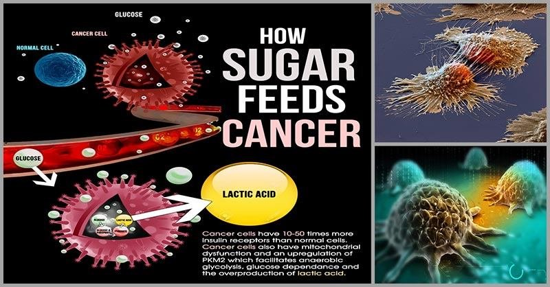
ပုံမှန်ထက်အဆမတန်များသောသကြားဓါတ်များသည်ဆဲလ်များ၏အတွင်းပိုင်းသို့ရောက်ရှိသွားသောအခါဆဲလ်များ၏အတွင်းပိုင်းတွင်ရှိသော(mitochondria)ဟုခေါ်သောစွမ်းအင်ထုတ်ပေးသောဌာနတွင်၊နံပါတ်(၁)သကြားဓါတ်အလွန်များပြားလာပြီး၊နံပါတ်(၂)အောက်ဆီဂျင်မလုံလောက်ခြင်းဖြစ်လာပါမည်။ဤအခြေအနေနှစ်ရပ်ကြုံလာပြီဆိုလျင်စွမ်းအင်ထုတ်ပေးသောဌာန(mitochondria)သည်သကြားဓါတ်များကိုအမြန်ဖြုန်းတီးပြစ်နိုင်ရန်အတွက်အခြားရွေးချယ်စရာနည်းလမ်းမရှိတော့သဖြင့်ပုံမှန်ထက်သကြားဓါတ်သုံးစွဲမှု(၉)ဆပို၍များသောကင်ဆာဆဲလ်များကဲ့သိုသကြားဓါတ်များကိုအချည်ဖေါက်သုံးစွဲခြင်း(fermentation) ခေါ်ဆိုးရွားသောလုပ်ငန်းတစ်ခုကိုမလွဲသာမရှောင်သာ၍စတင်ပြုလုပ်ရပါတော့မည်။
——–
သကြားဓါတ်ကိုအချည်ဖေါက်ခြင်းလုပ်ငန်းဆိုသည်မှာပုံမှန်ဆဲလ်များ၏ပုံမှန်လုပ်ငန်းစဉ်မဟုတ်တော့ပဲ၊ကင်ဆာဆဲလ်များ၏လုပ်ရိုးလုပ်စဉ်အလေ့အကျင့်တခုဖြစ်နေသဖြင့်၊ဤသို့အကြိမ်ကြိမ်ကျင့်သုံးပြုလုပ်မှုများပြားလာသောအခါ၊အလေ့အကျင့်တခုလိုဖြစ်သွားပြီး၊ထိုပုံမှန်ဆဲလ်များသည်(cancer gene expression)ဖြစ်စဉ်ဖြစ်ပေါ်လာကာ၊ကင်ဆာဆဲလ်များအဖြစ်သို့အလိုလိုမျိုးရိုးဗီဇပြောင်းလည်းသွားပါသဖြင့်အလွန်ဆိုးရွားသောအခြေအနေတခုသို့ရောက်ရှိသွားနိုင်ပါသည်။
——
ဆဲလ်များသည်ကင်ဆာဆဲလ်များအဖြစ်သို့အလိုလိုပြောင်းလဲသွားရခြင်းဖြစ်ပေါ်လာရုံတင်မကပဲ၊ပြောင်းလဲသွားသည်နှင့်ချက်ခြင်းပင်သကြားဓါတ်များအားအလုံအလောက်ဆက်လက်ရရှိနေသေးသဖြင့်၊(ဝါးဘတ်နိယာမ)၏အကျိုးသက်ရောက်မှု(Warburg’s effect)အရ၊ကင်ဆာဆဲလ်များသည်မည်သို့မျှထိန်းချုပ်၍မရနိုင်လောက်အောင်လျှင်မြန်စွာအကြီးအကျယ်တကိုယ်လုံးဆီသိုပြန့်ပွားသွားပါတော့မည်။နဂိုကပင်သကြားဓါတ်များပြားမှုကြောင့်ပုံမှန်အလုပ်မလုပ်နိုင်ပဲပျက်စီးနေသောမိမိ၏ခုခံအားစနစ်သည်လည်းကင်ဆာဆဲလ်များကိုလျှစ်လျူရှုုနေရုံမှလွဲ၍မည်သို့မှမတတ်နိုင်တော့ပေ။
—————-
(ဝါးဘတ်)ဆိုသူမှာ(၁၉၂၄)ခုနစ်ကသကြားဓာတ်ကြောင့်ကင်ဆာဆဲလ်များဖြစ်ပေါ်ပွားများလာရသည်။သကြားဓါတ်လုံလောက်စွာမရရှိပါကကင်ဆာဆဲလ်များဆက်လက်၍အသက်မရှင်နိုင်ပဲသေဆုံးသွားသည်။ကင်ဆာဆဲလ်များသည်အခြားပုံမှန်ဆဲလ်များကဲ့သို့အခြားသောစွမ်းအင်များကိုအသုံးမပြုနိုင်ပဲ၊သကြားဓါတ်တခုတည်းကိုသာလျှင်အချည်ဖေါက်ခြင်း(fermentation)နည်းဖြင့်စွမ်းအင်ထုတ်ယူကာရှင်သန်ပွားများနေရသည်ဆိုသော၊ကင်ဆာရောဂါကုသမှုအတွက်အလွန်အသုံးဝင်မည့်သဘာဝသဘောတရားတစ်ခုကိုကမ္ဘာပေါ်တွင်ပထမဦးဆုံးရှာဖွေတွေ့ရှိခဲ့သဖြင့်ဆေးပညာတွင်နိုဘယ်ဆုကိုဆွတ်ခူးရရှိခဲ့သောဂျာမန်ဆေးသိပ္ပံပညာရှင်(Otto Heinrich Warburg)ပင်ဖြစ်ပါသည်။
——–
သို့သော်အလွန်ဝမ်းနည်းစရာကောင်းသည်မှာယနေ့ခေတ် အရင်းရှင်ဆေးပညာသည်ထို(ဝါးဘတ်)၏နိုဘယ်ဆုပေးရလောက်အောင်ထူးခြားသောတွေ့ရှိချက်များကိုပြည်ဖုံးကားချ၍လူနာများအားရောဂါတကယ်မပျောက်ပဲမိမိတို့အကျိုးအမြတ်သာများစွာရရှိနိုင်မည့်ကင်ဆာကုထုံးနည်းလမ်းများကိုသာဆက်လက်၍အသုံးပြုနေကြခြင်းပင်ဖြစ်ပါသည်။လွန်ခဲ့သောနစ်တစ်ရားနီးပါးခန့်(၁၉၂၄)ခုနစ်ကတည်းကပေါ်ထွက်နေခဲ့သော(ဝါးဘတ်)နိယာမကိုစာဖတ်သူမိမိတို့မှမသိထားခြင်းမှာအရင်းရှင်များ၏အပြစ်လား၊မိမိတို့၏လေ့လာမှုအားနည်းခြင်းကြောင့်လား၊မိမိတို့ကအရင်းရှင်များ၊ဆရာဝန်များ၊ဆေးရုံဆေးခန်းများကိုအဆုံးမဲ့ယုံကြည်ခဲ့မိ၍မိမိတို့၏ကျန်းမာရေးကိစ္စကိုမိမိတို့ကိုယ်တိုင်မလေ့လာ၊မစူးစမ်းပဲ၊မိမိ၏အသက်နှင့်ခန္ဓာကိုပုံပြီးအပ်ထားခဲ့မိ၍လားဆိုသည်မှာမိမိသာလျင်အသိဆုံးဖြစ်ပါမည်။
———-
ဤဆောင်းပါးကိုဖတ်ရှု့ချင်းအားဖြင့်ရရှိလာသော၊သကြားဓါတ်သည်ကင်ဆာရောဂါကိုဖြစ်ပေါ်ပွားများလာစေသောအကြောင်းတရားတခုဖြစ်သည်ဆိုသောအသိပညာထူးသည်အဖိုးမဖြတ်နိုင်သောအသိထူးတစ်ခုဖြစ်သော်လည်းစာထဲတွင်သာထားလိုက်ပြီး၊မိမိကိုယ်တိုင်ချက်ချင်းမလိုက်နာမကျင့်သုံးလျင်၊ကင်ဆာဆဲလ်များကသာလက်ဦးမှုရရှိသွားမည်ဖြစ်ပါသည်။သကြားဓာတ်သည်ကင်ဆာဆဲလ်များ၏အဓိကအဟာရဖြစ်သဖြင့်သကြားဓါတ်အားလုံးဝရှောင်ကြဉ်လိုက်သောနေထိုင်စားသောက်နည်း(low carb ketogenic diet)သည်ကင်ဆာရောဂါကိုသာမကပဲ၊ဆီးချိုရောဂါနှင့်အခြားသောရောဂါများကိုပါကာကွယ်နိုင်သဖြင့်ကမ္ဘာပေါ်တွင်အလွန်နာမည်ကြီး၍အလွန်အကျိုးများသောနည်းလမ်းတခုဖြစ်လာပါတော့သည်။
————
ကင်ဆာဆိုသည့်ရောဂါဆိုးဖြစ်လာလျှင်အမြစ်ပြတ်ပျောက်အောင်ကုသနိုင်သည့်ဆေးဆိုသည်မှာယနေ့ခေတ်ဆေးပညာတွင်မရှိသေးပါ။ကင်ဆာဖြစ်လာလျင်ခွဲစိတ်ကုသမှု့ခံရမည်။(Radiation Therapy)ဟုခေါ်သောအလွန်ပြင်းထန်သည့်ဓါတ်ရောင်ခြည်များနှင့်ကင်ပြီးကုသခြင်းခံရပါမည်၊ကီမိုသရဖီ(Chemotherapy)ဟုခေါ်သောအလွန်ကြောက်စရာကောင်းပြီးမိမိခန္ဒာကိုယ်အားလဲအဆိပ်သင့်စေကာမိမိ၏ခုခံအားစနစ်တစ်ခုလုံးကိုလဲပျက်စီးဆုံးရံှုးစေမည့်အဆိပ်ကျွေး၍ကုသသောကုသနည်းဖြင့်လဲကုသခြင်းခံရပါမည်။သို့သော်ပျောက်ကင်းမသွားပဲပို၍ဆိုးရွားလာကာဓါတ်ရောင်ခြည်နှင့်ကီမိုသရဖီတို့မှရရှိသည့်အဆိပ်အတောက်များကြောင့်မသေခင်တွင်လဲအလူးအလဲ၊အဝီစိငရဲမှာကဲ့သို့အလွန်ပြင်းထန်သည့်ဝေဒနာများကိုလည်းအလူးအလဲခံစားသွားရမည်ဖြစ်ပြီးမရှုမလှသေပွဲဝင်ကြရမည်ဖြစ်ပါသည်။အသေမလှတော့သဖြင့်ဘဝကူးလဲမကောင်းနိုင်တော့ပါ။
——–
ကြိုတင်ကာကွယ်ချင်းသည်ကုသခြင်းထက်ပို၍ထိရောက်၊အကျိုးရှိသဖြင့်၊မိမိမှာထိုကဲ့သို့သောအဖြစ်ဆိုးမျိုးမကြုံုလိုလျှင်(ဝါ)မဖြစ်လိုလျှင်၊ယခုချက်ချင်းပင်ဆေးပညာ၏နိုဘယ်ဆုရှင်(Otto Heinrich Warburg)၏သီအိုရီကိုအသုံးပြုပြီးသကြားဓါတ်(ကာဗိုဟိုက်ဒရိတ်)ပါသောအစားအသောက်များကိုကြိုတင်ရှောင်ကြဉ်၍နေထိုင်စားသောက်ရန်မှာအလွန်အရေးကြီးနေသည့်အပြင်(low carb ketogenic diet)ဟုခေါ်သောအလွန်အကျိုးကျေးဇူးများသောစားသောက်နေထိုင်နည်းစနစ်ကိုလဲစနစ်တကျလေ့လာသင်ယူထားရမည်ဖြစ်ပါသည်။ထိုနည်းအားအမြဲမကျင့်သုံးနိုင်လျင်လည်း၊မကြာမကြာထိုနည်းစနစ်ကိုကျင့်သုံးပေးခြင်းအားဖြင့်အလိုလိုကင်ဆာမှအစပြု၍ရောဂါအားလုံးဘေးရန်မှကာကွယ်နိုင်မည်ဖြစ်ပါသည်။ထိုနည်းစနစ်ကိုလည်းအများအကျိုးကိုမျော်ကိုး၍မကြာမီအကျယ်ရေးသားပေးပါဦးမည်။
———
လိုရင်းကိုပြန်၍ဆက်ပါဦးမည်။ဤကဲ့သို့သောအခြေအနေဆိုးများဖြစ်လာနိုင်သည်ကိုမိမိခန္ဓာကိုယ်ကကြိုတင်သိရှိနေသဖြင့်ဤအခြေအနေဆိုးများသို့ရောက်ရှိမသွားနိုင်စေရန်ကာကွယ်ပေးရမည့်တာဝန်မှာအသဲ၏တာဝန်သာလျှင်ဖြစ်လာပါတော့သည်။ထို့ကြောင့်သွေးထဲတွင်သကြားဓါတ်များလာသည်နှင့်အမျှအသဲကမိမိတွင်ရှိနေသောနေရာလွတ်များတွင်(ဂလိုင်ဂိုဂျင်အဖြစ်)ပထမဦးဆုံးသိုလှောင်ရပါသည်။သိုလှောင်မှုကလည်းကန့်သတ်ချက်အတိုင်းအတာအနည်းငယ်သာရှိသဖြင့်တစ်ခဏအတွင်းပြည့်သွားပါတော့သည်။ဤအချိန်တွင်အခြားရွေးစရာနည်းလမ်းမရှိတော့၍အသဲသည်သကြားဓါတ်များကိုအဆီအဖြစ်သို့ပြောင်းလဲခြင်းလုပ်ငန်း(hepatic de novo lipogenesis pathway)လမ်းကြောင်းကိုစတင်လုပ်ဆောင်ရပါတော့သည်။
———–
အသဲသည်သကြားဓါတ်များကိုအဆီအဖြစ်သို့ပြောင်းလဲပြီးလျှင်လည်းအဆီများကို(VLDL)ဟုခေါ်သောကော်လက်စထရောများ၏အကူအညီနှင့်ခန္ဓာကိုယ်အစိတ်အပိုင်းအသီးသီးတွင်သိုလှောင်ထားနိုင်ရန်ပို့ဆောင်ပေးဖို့ကြိုးစားရပြန်သည်။အသဲသည်သကြားဓါတ်များကိုအဆီအဖြစ်ပြောင်းလဲရုံတင်မကပဲခန္ဓာကိုယ်၏အစိတ်အပိုင်းများဆီသို့ပါသယ်ယူပို့ဆောင်ထုတ်ယူသွားပေးနိုင်ရန်(VLDL)ဟုခေါ်သောကော်လက်စထရောများကိုလည်းဆိုင်းငံ့မှုမရှိပဲလျင်မြန်စွာထုတ်လုပ်ပေးရပါတော့သည်။
——–
အကယ်၍သကြားဓါတ်အားလုံးကိုအသဲကအဆီအဖြစ်အောင်မြင်စွာပြောင်းလည်းပြစ်နိုင်ပြီး၊(VLDL)ခေါ်ကော်လက်စထရောများကိုလည်းအလုံအလောက်ထုတ်လုပ်ပေးနိုင်ကာ၊ထို(VLDL)ကော်လက်စထရောများ၏အကူအညီဖြင့်ခန္ဓာကိုယ်၏အစိတ်အပိုင်းအသီးသီးဆီသို့အောင်မြင်စွာသယ်ယူပို့ဆောင်သွားပြီးသိုလှောင်ထားလိုက်နိုင်မည်ဆိုပါကဆီးချိုရောဂါအမှတ်(၂)ဆိုသည်မှာဖြစ်လာစရာအကြောင်းမရှိတော့ပေ။ခန္ဓာကိုယ်အစိတ်အပိုင်းများတွင်တော့အဆီများစုဝေးများပြားလာကာကိုယ်အလေးချိန်တော့တက်လာမည်ဖြစ်ပါသည်။
——-
သို့သော်လက်တွေ့တွင်ထိုသို့မျော်လင့်ထားသည့်အတိုင်းဖြစ်မလာနိုင်ခဲ့ပါ။သကြားဓါတ်မှအဆီများအဖြစ်သို့ပြောင်းလည်းပြစ်လိုက်နိုင်သည်ကတော့မှန်ပါသည်၊သို့သော်အကြောင်းများစွာမညီညွတ်သဖြင့်ဤပြောင်းလဲပြီးသွားသောအဆီများကိုခန္ဓာကိုယ်အစိတ်အပိုင်းအသီးသီးသို့အောင်မြင်စွာသယ်ထုတ်မသွားနိုင်ခဲ့ပါ။ဤအဆီများသည်အသဲထဲမှာသာလျှင်စုပုံကျန်ရစ်နေပြီး၊အချိန်ကြာလာသည်နှင့်အမျှအသဲထဲတွင်ပြည့်နှက်လာပါတော့သည်။အသဲတခုလုံးအဆီများဖုံးလွမ်းလာကာပုံမှန်အလုပ်မလုပ်နိုင်တော့ပဲအသဲအဆီဖုံးရောဂါ(Fatty Liver Disease)ဖြစ်ပေါ်လာရပါတော့သည်။
———
အဘယ်သို့သောအကြောင်းတရားများကြောင့်အသဲသည်သကြားဓါတ်မှပြောင်းလဲပြီးသွားသောအဆီသစ်များကိုမိမိနှင့်ဝေးရာသို့သယ်ယူပို့ဆောင်မှုလုပ်ငန်းအားမပြုလုပ်မဆောင်ရွက်နိုင်ပဲမိမိကိုယ်တိုင်ပင်ထိခိုက်ပျက်စီးသွားရသနည်းဟုမေးလာပါက၊ထိုပြဿနာဖြစ်ပေါ်လာရခြင်း၏အကြောင်းတရားများကိုအသေးစိတ်သိရှိနားလည်နိုင်ရန်နံပါတ်(၂)လမ်းကြောင်းကြီးဖြစ်သောအသဲ၏(hepatic de novo lipogenesis pathway)ဟုခေါ်သောလမ်းကြောင်းနောက်တခုကိုနောက်ထပ်အပိုင်းများတွင်ဆက်လက်ပြီးရေးသားဖေါ်ပြသွားမည်ဖြစ်ပါသည်။ယခုတော့နားပါဦးမယ်။အားလုံးအနာရောဂါကင်းရှင်း၍ကျန်းမာကြပါစေ။
——
Kelvin Albert Power
(Nutrition Specialist, Florida, USA)
———–
(မှတ်ချက်။)ရှေ့အပိုင်းဆက်များတွင်ရေးသားထားသည့်ဆိုလိုရင်းများမှာဆီးချိုရောဂါရှိသူများသည်သကြားဓါတ်မမြင့်မားအောင်မထိန်းချုပ်သင့်ဟု(လုံးဝ)လုံးဝမဆိုလိုပါ။သကြားဓါတ်အားပုံမှန်ဖြစ်အောင်ဦးစွာပထမထိန်းချုပ်ရမည်ဖြစ်ပါသည်။သကြားဓါတ်ထိန်းချုပ်ရာတွင်လဲဆေးများထက်သဘာဝနည်းလမ်းများကိုပို၍အားကိုးရမည်ဖြစ်ပါသည်။သို့သော်သကြားဓါတ်ထိန်းချုပ်ထားရုံနှင့်မိမိတို့ကိစ္စပြီးဆုံးမသွားပဲအောက်ခံရောဂါဖြစ်ပွားရသောအကြောင်းတရားများကိုလည်းအဓိကထားရှာဖွေ၍ပြုပြင်ကုသကြရမည်ဟုဆိုလိုထားခြင်းဖြစ်ပါသည်။
———- REFERENCES————–
1. Xu XD, Shao SX, Jiang HP, Cao YW, Wang YH, Yang XC, Wang YL, Wang XS, Niu HT. Warburg effect or reverse Warburg effect? A review of cancer metabolism. Oncol Res Treat. 2015;38:117–22. [PubMed] [Google Scholar]
2. Dang CV. Links between metabolism and cancer. Genes Dev. 2012;26:877–90. [PMC free article] [PubMed] [Google Scholar]
3. Ward PS, Thompson CB. Metabolic reprogramming: a cancer hallmark even warburg did not anticipate. Cancer Cell. 2012;21:297–308. [PMC free article] [PubMed] [Google Scholar]
4. Soga T. Cancer metabolism: key players in metabolic reprogramming. Cancer Sci. 2013;104:275–81. [PubMed] [Google Scholar]
5. Bayley JP, Devilee P. The Warburg effect in 2012. Curr Opin Oncol. 2012;24:62–7. [PubMed] [Google Scholar]
6. Koppenol WH, Bounds PL, Dang CV. Otto Warburg’s contributions to current concepts of cancer metabolism. Nat Rev Cancer. 2011;11:325–37. [PubMed] [Google Scholar]
7. Schuurbiers OC, Meijer TW, Kaanders JH, Looijen-Salamon MG, de Geus-Oei LF, van der Drift MA, van der Heijden EH, Oyen WJ, Visser EP, Span PN, Bussink J. Glucose Metabolism in NSCLC Is Histology-Specific and Diverges the Prognostic Potential of 18FDG-PET for Adenocarcinoma and Squamous Cell Carcinoma. J Thorac Oncol. 2014;9:1485–93. [PubMed] [Google Scholar]
8. Zeiss K, Parhofer KG, Heinemann V, Haas M, Laubender RP, Holdenrieder S, Schulz C, Boeck S. Glucose and lipid metabolism in patients with advanced pancreatic cancer receiving palliative chemotherapy. Anticancer Res. 2013;33:287–92. [PubMed] [Google Scholar]
9. Chen X, Qian Y, Wu S. The Warburg Effect: Evolving Interpretations Of An Established Concept. Free Radic Biol Med. 2015;79:253–63. [PMC free article] [PubMed] [Google Scholar]
10. Held-Warmkessel J, Dell DD. Lactic acidosis in patients with cancer. Clin J Oncol Nurs. 2014;18:592–4. [PubMed] [Google Scholar]
11. Shiraishi T, Verdone JE, Huang J, Kahlert UD, Hernandez JR, Torga G, Zarif JC, Epstein T, Gatenby R, McCartney A, Elisseeff JH, Mooney SM, An SS, Pienta KJ. Glycolysis is the primary bioenergetic pathway for cell motility and cytoskeletal remodeling in human prostate and breast cancer cells. Oncotarget. 2015;6:130–43. doi: 10.18632/oncotarget.2766. [PMC free article] [PubMed] [CrossRef] [Google Scholar]
12. Peppicelli S, Bianchini F, Calorini L. Extracellular acidity, a “reappreciated” trait of tumor environment driving malignancy: perspectives in diagnosis and therapy. Cancer Metastasis Rev. 2014;33:823–32. [PubMed] [Google Scholar]
13. Chen JQ, Russo J. Dysregulation of glucose transport, glycolysis, TCA cycle and glutaminolysis by oncogenes and tumor suppressors in cancer cells. Biochim Biophys Acta. 2012;1826:370–84. [PubMed] [Google Scholar]
14. Dang CV. Therapeutic targeting of Myc-reprogrammed cancer cell metabolism. Cold Spring Harb Symp Quant Biol. 2011;76:369–74. [PubMed] [Google Scholar]
15. Xia Y, Shen S, Verma IM. NF-κB, an active player in human cancers. Cancer Immunol Res. 2014;2:823–30. [PMC free article] [PubMed] [Google Scholar]
16. Lai L, Yan L, Gao S, Hu CL, Ge H, Davidow A, Park M, Bravo C, Iwatsubo K, Ishikawa Y, Auwerx J, Sinclair DA, Vatner SF, Vatner DE. Type 5 adenylyl cyclase increases oxidative stress by transcriptional regulation of manganese superoxide dismutase via the SIRT1/FoxO3a pathway. Circulation. 2013;127:1692–701. [PMC free article] [PubMed] [Google Scholar]
17. Kim JH, Qu A, Reddy JK, Gao B, Gonzalez FJ. Hepatic oxidative stress activates the Gadd45b gene via degradation of the transcriptional repressor STAT3. Hepatology. 2014;59:695–704. [PMC free article] [PubMed] [Google Scholar]
18. Keller KE, Tan IS, Lee YS. SAICAR stimulates pyruvate kinase isoform M2 and promotes cancer cell survival in glucose-limited conditions. Science. 2012;338:1069–72. [PMC free article] [PubMed] [Google Scholar]
19. Pflaum J, Schlosser S, Müller M. p53 Family and Cellular Stress Responses in Cancer. Front Oncol. 2014;4:285. [PMC free article] [PubMed] [Google Scholar]
20. Vigneron A, Vousden KH. p53, ROS and senescence in the control of aging. Aging (Albany NY) 2010;2:471–4. doi: 10.18632/aging.100189. [PMC free article] [PubMed] [CrossRef] [Google Scholar]
21. Migliaccio E, Giorgio M, Pelicci PG. p53 and aging: role of p66Shc. Aging (Albany NY) 2013;5:488–9. doi: 10.18632/aging.100583. [PMC free article] [PubMed] [CrossRef] [Google Scholar]
22. Blagosklonny MV. Tumor suppression by p53 without apoptosis and senescence: conundrum or rapalog-like gerosuppression? Aging (Albany NY) 2012;4:450–5. doi: 10.18632/aging.100475. [PMC free article] [PubMed] [CrossRef] [Google Scholar]
23. Guo H, Liu Z, Xu B, Hu H, Wei Z, Liu Q, Zhang X, Ding X, Wang Y, Zhao M, Gong Y, Shao C. Chemokine receptor CXCR2 is transactivated by p53 and induces p38-mediated cellular senescence in response to DNA damage. Aging Cell. 2013;12:1110–21. [PubMed] [Google Scholar]
24. Johnson RF, Perkins ND. Nuclear factor-κB, p53, and mitochondria: regulation of cellular metabolism and the Warburg effect. Trends Biochem Sci. 2012;37:317–24. [PubMed] [Google Scholar]
25. Broek RV, Mohan S, Eytan DF, Chen Z, Van Waes C. The PI3K/Akt/mTOR axis in head and neck cancer: functions, aberrations, crosstalk, and therapies. Oral Dis. 2015;21:815–25. [PubMed] [Google Scholar]
26. Liao JM, Cao B, Zhou X, Lu H. New insights into p53 functions through its target microRNAs. J Mol Cell Biol. 2014;6:206–13. [PMC free article] [PubMed] [Google Scholar]
27. Wang SJ, Gu W. To be, or not to be: functional dilemma of p53 metabolic regulation. Curr Opin Oncol. 2014;26:78–85. [PMC free article] [PubMed] [Google Scholar]
28. Madan E, Gogna R, Bhatt M, Pati U, Kuppusamy P, Mahdi AA. Regulation of glucose metabolism by p53: emerging new roles for the tumor suppressor. Oncotarget. 2011;2:948–57. doi: 10.18632/oncotarget.389. [PMC free article] [PubMed] [CrossRef] [Google Scholar]
29. Zhang XD, Qin ZH, Wang J. The role of p53 in cell metabolism. Acta Pharmacol Sin. 2010;31:1208–12. [PMC free article] [PubMed] [Google Scholar]
30. Zhang C, Liu J, Wu R, Liang Y, Lin M, Liu J, Chan CS, Hu W, Feng Z. Tumor suppressor p53 negatively regulates glycolysis stimulated by hypoxia through its target RRAD. Oncotarget. 2014;5:5535–46. doi: 10.18632/oncotarget.2137. [PMC free article] [PubMed] [CrossRef] [Google Scholar]
31. Aquilano K, Baldelli S, Pagliei B, Cannata SM, Rotilio G, Ciriolo MR. p53 orchestrates the PGC-1α-mediated antioxidant response upon mild redox and metabolic imbalance. Antioxid Redox Signal. 2013;18:386–99. [PMC free article] [PubMed] [Google Scholar]
32. Rajeshkumar NV, Dutta P, Yabuuchi S, de Wilde RF, Martinez GV, Le A, Kamphorst JJ, Rabinowitz JD, Jain SK, Hidalgo M, Dang CV, Gillies RJ, Maitra A. Therapeutic Targeting of the Warburg Effect in Pancreatic Cancer Relies on an Absence of p53 Function. Cancer Res. 2015;75:3355–64. [PMC free article] [PubMed] [Google Scholar]
33. Kim SJ, Jung HJ, Lim CJ. Reactive Oxygen Species-Dependent Down-Regulation of Tumor Suppressor Genes PTEN, USP28, DRAM, TIGAR, and CYLD Under Oxidative Stress. Biochem Genet. 2013;51:901–15. [PubMed] [Google Scholar]
34. Desideri E, Vegliante R, Ciriolo MR. Mitochondrial dysfunctions in cancer: Genetic defects and oncogenic signaling impinging on TCA cycle activity. Cancer Lett. 2015;356:217–23. [PubMed] [Google Scholar]
35. Liu J, Zhang C, Lin M, Zhu W, Liang Y, Hong X, Zhao Y, Young KH, Hu W, Feng Z. Glutaminase 2 negatively regulates the PI3K/AKT signaling and shows tumor suppression activity in human hepatocellular carcinoma. Oncotarget. 2014;5:2635–47. doi: 10.18632/oncotarget.1862. [PMC free article] [PubMed] [CrossRef] [Google Scholar]
36. Mucaj V, Shay JE, Simon MC. Effects of hypoxia and HIFs on cancer metabolism. Int J Hematol. 2012;95:464–70. [PubMed] [Google Scholar]
37. Seton-Rogers S. Hypoxia: HIF switch. Nat Rev Cancer. 2011;11:391. [PubMed] [Google Scholar]
38. Leontieva OV, Natarajan V, Demidenko ZN, Burdelya LG, Gudkov AV, Blagosklonny MV. Hypoxia suppresses conversion from proliferative arrest to cellular senescence. Proc Natl Acad Sci U S A. 2012;109:13314–8. [PMC free article] [PubMed] [Google Scholar]
39. Nardinocchi L, Puca R, D’Orazi G. HIF-1α antagonizes p53-mediated apoptosis by triggering HIPK2 degradation. Aging (Albany NY) 2011;3:33–43. doi: 10.18632/aging.100254. [PMC free article] [PubMed] [CrossRef] [Google Scholar]
40. Meijer TW, Kaanders JH, Span PN, Bussink J. Targeting hypoxia, HIF-1, and tumor glucose metabolism to improve radiotherapy efficacy. Clin Cancer Res. 2012;18:5585–94. [PubMed] [Google Scholar]
41. Ahn GO, Seita J, Hong BJ, Kim YE, Bok S, Lee CJ, Kim KS, Lee JC, Leeper NJ, Cooke JP, Kim HJ, Kim IH, Weissman IL, Brown JM. Transcriptional activation of hypoxia-inducible factor-1 (HIF-1) in myeloid cells promotes angiogenesis through VEGF and S100A8. Proc Natl Acad Sci U S A. 2014;111:2698–703. [PMC free article] [PubMed] [Google Scholar]
42. Kim KJ, Choi JS, Kang I, Kim KW, Jeong CH, Jeong JW. Melatonin suppresses tumor progression by reducing angiogenesis stimulated by HIF-1 in a mouse tumor model. J Pineal Res. 2013;54:264–70. [PubMed] [Google Scholar]
43. Zwaans BM, Lombard DB. Interplay between sirtuins, MYC and hypoxia-inducible factor in cancer-associated metabolic reprogramming. Dis Model Mech. 2014;7:1023–32. [PMC free article] [PubMed] [Google Scholar]
44. Denko NC. Hypoxia, HIF1 and glucose metabolism in the solid tumour. Nat Rev Cancer. 2008;8:705–13. [PubMed] [Google Scholar]
45. Darnell JE., Jr STAT3, HIF-1, glucose addiction and Warburg effect. Aging (Albany NY) 2010;2:890–1. doi: 10.18632/aging.100239. [PMC free article] [PubMed] [CrossRef] [Google Scholar]
46. Yang C, Jiang L, Zhang H, Shimoda LA, DeBerardinis RJ, Semenza GL. Analysis of hypoxia-induced metabolic reprogramming. Methods Enzymol. 2014;542:425–55. [PubMed] [Google Scholar]
47. Semenza GL. HIF-1 mediates metabolic responses to intratumoral hypoxia and oncogenic mutations. J Clin Invest. 2013;123:3664–71. [PMC free article] [PubMed] [Google Scholar]
48. Starska K, Forma E, Jóźwiak P, Bryś M, Lewy-Trenda I, Brzezińska-Błaszczyk E, Krześlak A. Gene and protein expression of glucose transporter 1 and glucose transporter 3 in human laryngeal cancer-the relationship with regulatory hypoxia-inducible factor-1α expression, tumor invasiveness, and patient prognosis. Tumour Biol. 2015;36:2309–21. [PMC free article] [PubMed] [Google Scholar]
49. Wolf A, Agnihotri S, Micallef J, Mukherjee J, Sabha N, Cairns R, Hawkins C, Guha A. Hexokinase 2 is a key mediator of aerobic glycolysis and promotes tumor growth in human glioblastoma multiforme. J Exp Med. 2011;208:313–26. [PMC free article] [PubMed] [Google Scholar]
50. Cheng SC, Quintin J, Cramer RA, Shepardson KM, Saeed S, Kumar V, Giamarellos-Bourboulis EJ, Martens JH, Rao NA, Aghajanirefah A, Manjeri GR, Li Y, Ifrim DC, et al. mTOR- and HIF-1α-mediated aerobic glycolysis as metabolic basis for trained immunity. Science. 2014;345:1250684. [PMC free article] [PubMed] [Google Scholar]
51. Hussien R, Brooks GA. Mitochondrial and plasma membrane lactate transporter and lactate dehydrogenase isoform expression in breast cancer cell lines. Physiol Genomics. 2011;43:255–64. [PMC free article] [PubMed] [Google Scholar]
52. Zhao L, Yang YF, Gao YB, Wang SM, Wang LF, Zuo HY, Dong J, Xu XP, Su ZT, Zhou HM, Zhu LL, Peng RY. Upregulation of HIF-1α Via Activation of ERK and PI3K Pathway Mediated Protective Response to Microwave-Induced Mitochondrial Injury in Neuron-Like Cells. Mol Neurobiol. 2014;50:1024–34. [PubMed] [Google Scholar]
53. Starska K, Forma E, Jóźwiak P, Bryś M, Lewy-Trenda I, Brzezińska-Błaszczyk E, Krześlak A. Gene and protein expression of glucose transporter 1 and glucose transporter 3 in human laryngeal cancer-the relationship with regulatory hypoxia-inducible factor-1α expression, tumor invasiveness, and patient prognosis. Tumour Biol. 2015;36:2309–21. [PMC free article] [PubMed] [Google Scholar]
54. Chen JQ, Russo J. Dysregulation of glucose transport, glycolysis, TCA cycle and glutaminolysis by oncogenes and tumor suppressors in cancer cells. Biochim Biophys Acta. 2012;1826:370–84. [PubMed] [Google Scholar]
55. Ong SG, Lee WH, Theodorou L, Kodo K, Lim SY, Shukla DH, Briston T, Kiriakidis S, Ashcroft M, Davidson SM, Maxwell PH, Yellon DM, Hausenloy DJ. HIF-1 reduces ischaemia-reperfusion injury in the heart by targeting the mitochondrial permeability transition pore. Cardiovasc Res. 2014;104:24–36. [PubMed] [Google Scholar]
56. Zhao T, Zhu Y, Morinibu A, Kobayashi M, Shinomiya K, Itasaka S, Yoshimura M, Guo G, Hiraoka M, Harada H. HIF-1-mediated metabolic reprogramming reduces ROS levels and facilitates the metastatic colonization of cancers in lungs. Sci Rep. 2014;4:3793. [PMC free article] [PubMed] [Google Scholar]
57. Xie JM, Li B, Yu HP, Gao QG, Li W, Wu HR, Qin ZH. TIGAR has a dual role in cancer cell survival through regulating apoptosis and autophagy. Cancer Res. 2014;74:5127–38. [PubMed] [Google Scholar]
58. Madan E, Gogna R, Kuppusamy P, Bhatt M, Pati U, Mahdi AA. TIGAR induces p53-mediated cell-cycle arrest by regulation of RB-E2F1 complex. Br J Cancer. 2012;107:516–26. [PMC free article] [PubMed] [Google Scholar]
59. Peña-Rico MA, Calvo-Vidal MN, Villalonga-Planells R, Martínez-Soler F, Giménez-Bonafé P, Navarro-Sabaté À, Tortosa A, Bartrons R, Manzano A. TP53 induced glycolysis and apoptosis regulator (TIGAR) knockdown results in radiosensitization of glioma cells. Radiother Oncol. 2011;101:132–9. [PubMed] [Google Scholar]
60. Wanka C, Steinbach JP, Rieger J. Tp53-induced glycolysis and apoptosis regulator (TIGAR) protects glioma cells from starvation-induced cell death by up-regulating respiration and improving cellular redox homeostasis. J Biol Chem. 2012;287:33436–46. [PMC free article] [PubMed] [Google Scholar]
61. Won KY, Lim SJ, Kim GY, Kim YW, Han SA, Song JY, Lee DK. Regulatory role of p53 in cancer metabolism via SCO2 and TIGAR in human breast cancer. Hum Pathol. 2012;43:221–8. [PubMed] [Google Scholar]
62. Gerin I, Noël G, Bolsée J, Haumont O, Van Schaftingen E, Bommer GT. Identification of TP53-induced glycolysis and apoptosis regulator (TIGAR) as the phosphoglycolate-independent 2,3-bisphosphoglycerate phosphatase. Biochem J. 2014;458:439–48. [PubMed] [Google Scholar]
63. Bolaños JP. TIGAR’s promiscuity. Biochem J. 2014;458:e5–7. [PubMed] [Google Scholar]
64. Ye L, Zhao X, Lu J, Qian G, Zheng JC, Ge S. Knockdown of TIGAR by RNA interference induces apoptosis and autophagy in HepG2 hepatocellular carcinoma cells. Biochem Biophys Res Commun. 2013;437:300–6. [PubMed] [Google Scholar]
65. Li M, Sun M, Cao L, Gu JH, Ge J, Chen J, Han R, Qin YY, Zhou ZP, Ding Y, Qin ZH. A TIGAR-regulated metabolic pathway is critical for protection of brain ischemia. J Neurosci. 2014;34:7458–71. [PubMed] [Google Scholar]
66. Cheung EC, Ludwig RL, Vousden KH. Mitochondrial localization of TIGAR under hypoxia stimulates HK2 and lowers ROS and cell death. Proc Natl Acad Sci U S A. 2012;109:20491–6. [PMC free article] [PubMed] [Google Scholar]
67. Lui VW, Wong EY, Ho K, Ng PK, Lau CP, Tsui SK, Tsang CM, Tsao SW, Cheng SH, Ng MH, Ng YK, Lam EK, Hong B, Lo KW, Mok TS, Chan AT, Mills GB. Inhibition of c-Met downregulates TIGAR expression and reduces NADPH production leading to cell death. Oncogene. 2011;30:1127–34. [PMC free article] [PubMed] [Google Scholar]
68. Dang CV. Cancer Cell Metabolism: There Is No ROS for the Weary. Cancer Discov. 2012;2:304–7. [PubMed] [Google Scholar]
69. Trejo-Solís C, Jimenez-Farfan D, Rodriguez-Enriquez S, Fernandez-Valverde F, Cruz-Salgado A, Ruiz-Azuara L, Sotelo J. Copper compound induces autophagy and apoptosis of glioma cells by reactive oxygen species and JNK activation. BMC Cancer. 2012;12:156. [PMC free article] [PubMed] [Google Scholar]
70. Yin L, Kufe T, Avigan D, Kufe D. Targeting MUC1-C is synergistic with bortezomib in downregulating TIGAR and inducing ROS-mediated myeloma cell death. Blood. 2014;123:2997–3006. [PMC free article] [PubMed] [Google Scholar]
71. Kim SJ, Jung HJ, Lim CJ. Reactive oxygen species-dependent down-regulation of tumor suppressor genes PTEN, USP28, DRAM, TIGAR, and CYLD under oxidative stress. Biochem Genet. 2013;51:901–15. [PubMed] [Google Scholar]
72. Pasquinelli AE. MicroRNAs and their targets: recognition, regulation and an emerging reciprocal relationship. Nat Rev Genet. 2012;13:271–82. [PubMed] [Google Scholar]
73. Rottiers V, Näär AM. MicroRNAs in metabolism and metabolic disorders. Nat Rev Mol Cell Biol. 2012;13:239–50. [PMC free article] [PubMed] [Google Scholar]
74. Kasinski AL, Slack FJ. Epigenetics and genetics. MicroRNAs en route to the clinic: progress in validating and targeting microRNAs for cancer therapy. Nat Rev Cancer. 2011;11:849–64. [PMC free article] [PubMed] [Google Scholar]
75. Chen B, Li H, Zeng X, Yang P, Liu X, Zhao X, Liang S. Roles of microRNA on cancer cell metabolism. J Transl Med. 2012;10:228. [PMC free article] [PubMed] [Google Scholar]
76. Gao P, Sun L, He X, Cao Y, Zhang H. MicroRNAs and the Warburg Effect: new players in an old arena. Curr Gene Ther. 2012;12:285–91. [PubMed] [Google Scholar]
77. Tomaselli S, Panera N, Gallo A, Alisi A. Circulating miRNA profiling to identify biomarkers of dysmetabolism. Biomark Med. 2012;6:729–42. [PubMed] [Google Scholar]
78. Singh PK, Brand RE, Mehla K. MicroRNAs in pancreatic cancer metabolism. Nat Rev Gastroenterol Hepatol. 2012;9:334–44. [PMC free article] [PubMed] [Google Scholar]
79. Fang R, Xiao T, Fang Z, Sun Y, Li F, Gao Y, Feng Y, Li L, Wang Y, Liu X, Chen H, Liu XY, Ji H. miR-143 regulates cancer glycolysis via targeting hexokinase 2. J Biol Chem. 2012;287:23227–35. [PMC free article] [PubMed] [Google Scholar]
80. Yao M, Wang X, Tang Y, Zhang W, Cui B, Liu Q, Xing L. Dicer mediating the expression of miR-143 and miR-155 regulates hexokinase II associated cellular response to hypoxia. Am J Physiol Lung Cell Mol Physiol. 2014;307:L829–37. [PubMed] [Google Scholar]
81. Alfarouk KO, Verduzco D, Rauch C, et al. Glycolysis, tumor metabolism, cancer growth and dissemination. A new pH-based etiopathogenic perspective and therapeutic approach to an old cancer question. Oncol Sci. 2014;1:777–91. [PMC free article] [PubMed] [Google Scholar]
82. Altenberg B, Greulich KO. Genes of glycolysis are ubiquitously over expressed in cancer classes. Genomics. 2004;84:1014–20. [PubMed] [Google Scholar]
83. Amoedo ND, Valencia JP, Rodrigues MF, Galina A, Rumjanek FD. How does the metabolism of tumor cells differ from that of normal cells? Biosci Rep. 2013;33:865–871. [PMC free article] [PubMed] [Google Scholar]
84. Anastasiou D, Poulogiannis G, Asara JM, et al. Inhibition of pyruvate kinase m2 by reactive oxygen species contributes to cellular antioxidant responses. Science. 2011;334:1278–83. [PMC free article] [PubMed] [Google Scholar]
85. Annibaldi A, Widmann C. Glucose metabolism in cancer cells. Curr Opin Clin Nutr Metab Care. 2011;13:466–70. [PubMed] [Google Scholar]
86. Baeriswyl V, Christofori G. The angiogenic switch in carcinogenesis. Semin Cancer Biol. 2009;19:329–37. [PubMed] [Google Scholar]
87. Barthel A, Okino ST, Liao J, et al. Regulation of GLUT1 gene transcription by the serine/threonine kinaseakt1. J Biol Chem. 1999;274:20281–86. [PubMed] [Google Scholar]
88. Bensaad K, Tsuruta A, Selak MA, et al. TIGAR, a p53-inducible regulator of glycolysis and apoptosis. Cell. 2006;126:107–20. [PubMed] [Google Scholar]
89. Berg JM, Tymoczko Jl, Stryer L Biochemistry. Glycolysis and Gluconeogenesis. 5th Edition. Chapter 16:2. New York: W H Freeman; 2002. pp. 634–5. [Google Scholar]
90. Blackwood EM, Kretzner L, Eisenman RN. Myc and Max function as a nucleoprotein complex. Curr Opin Gen Dev. 1992;2:227–35. [PubMed] [Google Scholar]
91. Boidot R, Vegran F, Meulle A, et al. Regulation of monocarboxylate transporter mct1 expression by p53 mediates inward and outward lactate fluxes in tumors. Cancer Res. 2012;72:939–48. [PubMed] [Google Scholar]
92. Bruick RK, McKnight SL. A conserved family of prolyl-4- hydroxylases that modify HIF. Science. 2001;294:133–740. [PubMed] [Google Scholar]
93. Burt MB, Hummy JL, Kooby DA, et al. Using positron emission tomography with [18F] FDG to predict tumor behavior in experimental colorectal cancer 1. Neo, 3. 2001:189–95. [PMC free article] [PubMed] [Google Scholar]
94. Buzzai M, Bauer DE, Jones RG, et al. The glucose dependence of Akt-transformed cells can be reversed by pharmacologic activation of fatty acid b-oxidation. Oncogene. 2005;24:4165–73. [PubMed] [Google Scholar]
95. Bykov VJ, Issaeva N, Shilov A, et al. Restoration of the tumor suppressor function to mutant p53 by a low-molecular-weight compound. Nat Med. 2002;8:282–8. [PubMed] [Google Scholar]
96. Cairns RA, Harris IS, Mak TW. Regulation of cancer cell metabolism. Nat Rev Cancer. 2011;11:85–95. [PubMed] [Google Scholar]
97. Cantley LC. The phosphoinositide 3-kinase pathway. Science. 2002;296:1655–57. [PubMed] [Google Scholar]
98. Chalhoub N, Baker SJ. PTEN and the PI3-kinase pathway in cancer. Annu Rev Pathol. 2009;4:127–500. [PMC free article] [PubMed] [Google Scholar]
99. Chaneton B, Hillmann P, Zheng L, et al. Serine is a natural ligand and allosteric activator of pyruvate kinase m2. Nat. 2012;491:458–462. [PMC free article] [PubMed] [Google Scholar]
100. Chen H, Liu H, Qing G. Targeting oncogenic Myc as a strategy for cancer treatment. Signal Transduct Target Ther. 2018;3:5. [PMC free article] [PubMed] [Google Scholar]
101. Chen JQ, Russo J. Dysregulation of glucose transport, glycolysis, TCA cycle and glutaminolysis by oncogenes and tumor suppressors in cancer cells. Biochim Biophys Acta. 2012;1826:370–84. [PubMed] [Google Scholar]
102. Chesney J, Mitchell R, Benigni F, et al. An inducible gene product for 6-phosphofructo-2-kinase with an AU-rich instability element:role in tumor cell glycolysis and the Warburg effect. Proc Natl Acad Sci U S A. 1999;96:3047–52. [PMC free article] [PubMed] [Google Scholar]
103. Cheung EC, Athineos D, Lee P, et al. TIGAR is required for efficient intestinal regeneration and tumorigenesis. Dev Cell. 2013;25:463–77. [PMC free article] [PubMed] [Google Scholar]
104. Chipuk JE, Kuwana T, Bouchier-Hayes L, et al. Direct activation of bax by p53 mediates mitochondrial membrane permeabilization and apoptosis. Science. 2004;303:1010–4. [PubMed] [Google Scholar]
105. Chow LM, Baker SJ. PTEN function in normal and neoplastic growth. Cancer Lit. 2006;241:184–196. [PubMed] [Google Scholar]
106. Clem B, Telang S, Clem A, et al. Small-molecule inhibition of 6-phosphofructo-2-kinase activity suppresses glycolytic flux and tumor growth. Mol Cancer Ther. 2008;7:110–20. [PubMed] [Google Scholar]